
Post Electrophoretic Analysis
Autoradiography
Autoradiography is the use of X-ray (or occasionally photographic) film to detect radioactive materials. It produces a permanent record of the positions and relative intensities of radiolabeled bands in a gel or blot. Typically, biomolecules are labeled with 32P or 35S, and detected by overnight film exposure. The table below gives the amounts of commonly used isotopes which can be detected by overnight autoradiography.
Autodiography Detection Limits
| Isotope | CPM Necessary for Detection | Energy per Emission (MEV) |
|---|---|---|
| 3H | >107 | 0.0055 |
| 14C | 2000 | 0.050 |
| 35S | 1000 | 0.167 |
| 32P | 100 | 0.70 |
| 125I | 10 | (gamma) |
Radioactive Exposure of Film
Beta particles emitted by radionuclides penetrate film emulsions to a depth proportional to their energy. As these particles pass through the film, they activate the silver halide crystals in the emulsion. Activated crystals are detected by reduction to black silver grains in the development step. The activated silver halide crystals are not stable, although they can be stabilized by further exposure to b-particles, or to light. On average, a crystal will require 5 "hits" from either radioactive emissions or light to be stably activated, and thus detected. In order to compensate for this instability, autoradiography is often carried out at -70°C, which stabilizes the activated crystals, enhancing sensitivity. Additional sensitivity is gained by "preflashing" the film before use. The film is exposed to a microsecond burst of light which is calibrated to bring the crystals in the film to a partially stabilized, activated state. Preflashed film requires only one "hit" per grain deposited. This not only increases sensitivity but also ensures that the film signal will be linear with the amount of radioactivity, even at very low signal levels.
Fluorography
The sensitivity of autoradiography can be greatly enhanced through the use of fluorography, which converts radioactive emissions into the light. Light penetrates film more efficiently than beta particles and is, therefore, more readily detected. Various phosphor compounds, such as National Diagnostics' Autofluor are available which can be dried into a gel, and absorb the energy from beta particles and re-emit it as light.
| Isotope | dpm/band | beq./band | Exposure (hours) |
|---|---|---|---|
| 3H | 500 | 8.3 | 48-72 |
| 3H | 5000 | 83 | 24 |
| 14C/35S | 300 | 5 | 24 |
| 14C/35S | 1000 | 17 | 8-12 |
For high-energy ß- particles, though, such as those from32P, the problem is not insufficient penetration but over-penetration, in which the emitted particles pass entirely through the film without activating the emulsion. To recover the energy of these "lost" particles, phosphorescent intensifying screens are used, which emit light on irradiation. Phosphorescent intensifying screens require low temperatures (-70°C) for optimum performance.
Autoradiography: The Procedure
Because water quenches beta particle emissions, optimum sensitivity is obtained using dried blots and gels. However, because drying blots permanently fixes the probe onto the membrane, if blots are to be stripped and re-probed, DO NOT DRY. Wrap in plastic wrap to prevent water and radioactivity from contacting the film. Note that 35S and 14C emit beta particles at low energy, and the majority of such particles are unable to penetrate a layer of plastic wrap. For 35S or 14C detection place the dry gel or blot directly on the film or use fluorographic enhancement. Use high-quality X-ray film: Kodak XAR or equivalent.
- Prepare gel or blot.
- Wrap wet samples in plastic wrap.
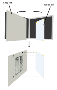
- Tape the sample to the inside face of an X-ray cassette, with enhancement screens for 32P if desired. It is essential to fasten the sample so that it does not move because it must later be aligned with the exposed film.
- Along one side, fasten a phosphorescent ruler or other orientation aid (radioactive dots). If a marker other than a ruler is used, be sure to include enough markers to unambiguously align the developed film to the sample.
- In the dark or under the safelight place one piece of X-ray film over the sample. For 35S or 14C, room temperature exposure is sufficient. For 32P, intensifying screens require a temperature of -70° C for exposure.
- Expose film. Use the tables above as a guide for the length of exposure. A good rule of thumb is that bands must be detectable on a Geiger counter for overnight exposure to be sufficient.
- To avoid condensation on the film allow the cassette to return to room temperature before opening. Develop the film according to the manufacturer's instructions.
NEXT TOPIC: Autoradiographic Enhancement with Autofluor