
Post Electrophoretic Analysis
Uneven Staining


Uneven staining is almost always the result of insufficient agitation during staining. The gel is initially more dense than the staining solution and will tend to sink to the bottom of the dish. Some portions of the gel will have stains under them, others will not, and the gel will show darker and lighter areas as a result.
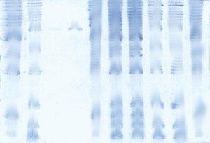
To recover this gel, simply continue the staining process with agitation (on a shaker), and the staining will become uniform.
- UV Shadowing
- Uneven Staining
- Staining Proteins Immobilized on Membranes
- Staining Protein Gels with Coomassie Blue
- Southern Blotting
- Smeared Bands
- Silver Staining Protein Gels
- Silver Staining DNA Gels
- Protein Fixation on Gels
- Post-Electrophoretic Visualization with Nuclistain
- Overview of Western Blotting
- Northern Blotting
- Method for Western Blotting
- Mechanism of Immunostaining
- Immunostaining with Alkaline Phosphatase
- Guide Strip Technique
- Faint bands, low background
- Faint Bands, High Background
- Ethidium Bromide Staining
- Enzyme Linked Immunosorbent Assay (ELISA)
- Coomassie Blue Stain- Troubleshooting
- Blotches on Gel
- Autoradiography
- Autoradiographic Enhancement with Autofluor
- An Overview of Northern and Southern Blotting
- Alkaline Blotting