Faint Bands, High Background
A high background which obscures the bands, or in combination with fainter than usual bands, indicates dye binding to the gel matrix, or contamination of the matrix with a dye-binding material (most often a protein).
Coomassie Blue R-250:
Destaining time too short: It takes hours for the dye to completely diffuse out of the gel after thorough staining. Early in the de-staining process, the background will be high and the bands weak. As the destaining progresses, bands will intensify somewhat as they capture free dye in the gel, and the background will decrease as dye elutes from the gel.
Protein contamination of the gel: This can occur during casting if the gel reagents are contaminated (most often by microbial growth), or during the run, if a sample of very high protein concentration is allowed to diffuse up into the running buffer. Once the upper tank running buffer is contaminated, the protein will be continuously fed into the gel, producing a diffuse background, rather than discrete bands. Use fresh, ultrapure gel and buffer reagents, and load only small volumes of high concentration samples.
Undissolved dye in staining solution: Dye crystals, if present, will deposit on the gel and dissolve into it, resulting in the high background that takes longer than usual to destain. Filter the dye solution to remove the undissolved dye.
ProtoBlue Safe/Colloidal Coomassie:
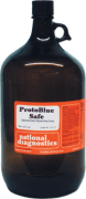
SDS in the gel: The primary cause of high background with Protoblue Safe is the presence of SDS in the gel. If the preliminary washes are not carried out properly, enough SDS will be carried into the staining solution to disrupt the colloidal dye suspension. As the colloidal particles break down, they allow the dye to diffuse into the gel. This background will eventually dissipate, but it can take overnight or longer to get a clear background. SDS will also inhibit the binding of the dye to the protein bands, further compromising sensitivity. Always use the completed pre-washing protocol.</.p>
Protein contamination of the gel: As with R-250 above, gels can be contaminated with protein if the reagents are old or impure, or if a sample of high protein concentration is allowed to diffuse into the upper running buffer. Use fresh, high-quality gel and buffer reagents, and load only small volumes of high concentration samples.