
Histology Fundamentals
Sectioning
Once embedded, tissues are cut into thin sections ready to be placed on a slide. This is done with a microtome, an apparatus for feeding the blocks past an ultrasharp blade with micron level precision.
Paraffin blocks can be sectioned with high-carbon steel blades. Plastic blocks (methacrylate, araldite, or epon) are sectioned with glass or diamond knives. A glass knife can section down to about 0.1 micron. Ultrathin sections for electron microscopy (below 100 nm) are best done with a diamond knife.
Sectioning tissues is an art. The selection of knife material, blade shape, cutting speed, knife angle and other variables must be determined through experience with the type of tissue and the particular equipment. Sections cut under non-optimal conditions will show tearing, ripping, "venetian blinds," holes, folding, etc. As sections are cut, they are floated on a warm water bath to smooth out any wrinkles. They are then picked up on a glass microscope slide.
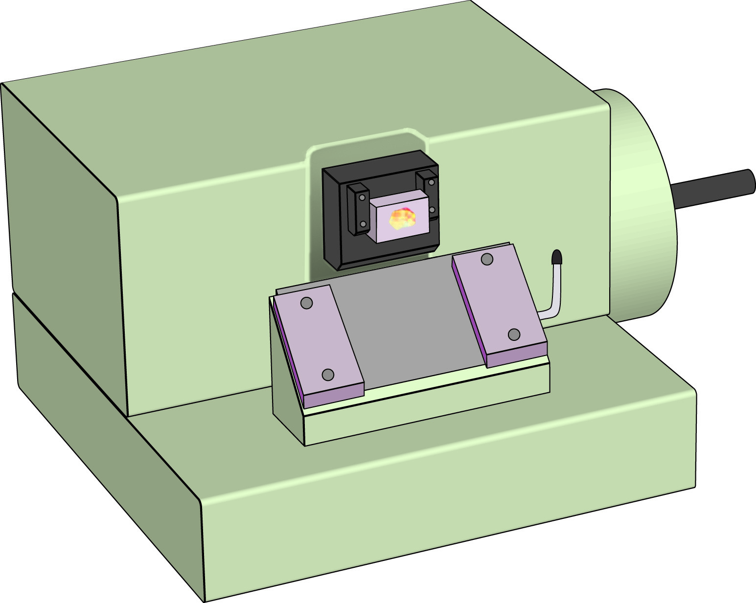
Rotary microtome for light microscopy.
The glass slides are then heated in a warm oven for about 15 minutes to help the section adhere to the slide. This step may be bypassed to preserve characteristics such as antigenicity. In this case, adhesive-coated slides may be substituted to pick up the sections. Typical adhesives for this purpose include starch, albumen, resins and combinations thereof. The adhered sections are then ready for further processing.
Frozen Sections
Sometimes in medical diagnosis it is necessary to perform a rapid analysis of a sample. This is facilitated by performing a frozen section. The piece(s) of tissue to be studied are snap frozen in a cold liquid or a cold environment (-20° to -70° Celsius). Freezing makes the tissue solid enough to section with a microtome.
Frozen sections are performed with an instrument called a cryostat, a refrigerated box containing a microtome. The temperature inside the cryostat is about -20° to -30° Celsius. The tissue sections are cut and picked up on a glass slide. The sections are then ready for staining.
NEXT TOPIC: Artifacts in Histological Sections