
Histology Fundamentals
Artifacts in Histologic Sections
Artifacts that appear in stained slides may result from a number of causes including improper fixation, the type of fixative, poor dehydration, improper reagents, or poor microtome sectioning.
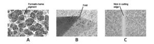
The presence of a fine black precipitate on the slides, often with no relationship to the tissue (i.e., the precipitate appears adjacent to tissues or within interstices or vessels) suggests the formation of formalin-heme pigment. The identification of this problem can be confirmed by polarized light microscopy. The pigment is birefringent in polarized light and will appear as numerous bright white motes on the slide. It forms when the formalin buffer is exhausted and the tissue becomes acidic, which promotes the formation of a complex of heme and formalin. Formalin-heme pigment is most often seen in tissues containing large amounts of blood or heme proteins, or in autopsy tissues. Tissues such as spleen and lymph node are particularly prone to this artifact. Making thin sections and using enough neutral-buffered formalin (10 to 1 ratio of fixative to tissue) will help. If the fixative solution in which the tissues are sitting is extremely murky brown to red, place the tissues in new fixative.
The presence of large irregular clumps of black precipitate on slides of tissues fixed in a mercurial fixative such as B-5 suggests that the tissues were not "dezenkerized" prior to staining. These black precipitates will also appear white with polarized light microscopy.
Tissues that are insufficiently dehydrated prior to clearing and infiltration with paraffin wax will be hard to section on the microtome, leading to tearing and holes in the sections. Tissue processor cycles should allow sufficient time for dehydration, and the final ethanol dehydrant solution should be at 100% concentration, which can be difficult to achieve in humid climates. Covering or sealing the solutions from ambient air will help. Air conditioning (with refrigerants, not with evaporative coolers) will also reduce humidity in the laboratory. As a clearing agent, toluene is more forgiving of poorly dehydrated tissues, but it is more expensive and presents more of a health hazard than most other clearing agents.
Though alcohols such as ethanol make excellent fixatives for cytologic smears, they tend to make tissue sections brittle, resulting in microtome sectioning artifacts with chattering and a 'venetian blind' appearance.
Bubbles under the coverslip may form when the mounting medium is too thin, and, as it dries, air is pulled in under the coverslip. Contamination of clearing agents or coverslipping media may also produce a bubbled appearance under the microscope. It is important to confirm that a clearing agent is compatible with the mounting medium to be used because some solvent may be carried over to the mounting stage.
NEXT TOPIC: The Chemistry of Dyes and Staining