Electrophoresis Articles
Overview of Western Blotting
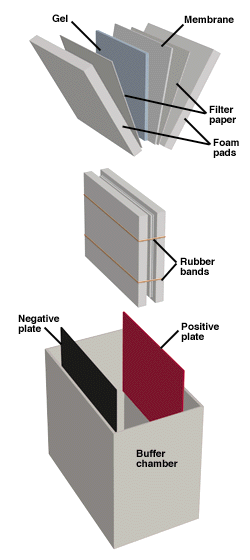
Proteins can also be detected immunologically following electrophoresis, a technique known as Western blotting. This method relies on the fact that most epitopes (sites recognized by antibodies, generally comprising several amino acids) are still recognizable following denaturing of the protein with SDS and binding to the surface of a membrane. The Western Blotting apparatus. Proteins…
Read MoreEnzyme Linked Immunosorbent Assay (ELISA)
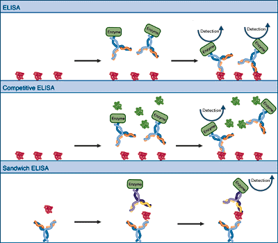
One of the most straightforward applications of immunological detection is the ELISA or enzyme-linked immunosorbent assay. In the simplest system, the bound antigen is probed with antibodies that carry covalently attached enzyme molecules. Antibody binding immobilizes the enzyme in the vicinity of the bound antigen, allowing detection of the antigen. Variations include a competition ELISA…
Read MoreImmunostaining with Alkaline Phosphatase
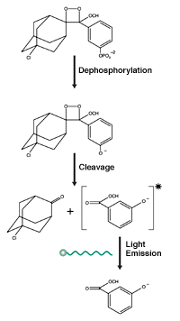
Alkaline phosphatase catalyzes the removal of a phosphate group from its substrate. A variety of synthetic substrates have been constructed which, on phosphate hydrolysis, liberate chromogens or luminescent compounds. A commonly used chromogenic substrate is bromochloroindoyl phosphate. (BCIP) in conjunction with nitro blue tetrazolium (NBT). Dephosphorylation of BCIP generates one half of an indigo dye…
Read MoreMechanism of Immunostaining
The basic method of immunostaining is to probe with an antibody that is bound to a detectable molecule such as an enzyme or a fluorescent dye. The highly specific binding interaction between antibodies and their unique antigens has been exploited to create sensitive and specific detection systems for proteins. An antibody can be raised and/or…
Read MoreStaining Proteins Immobilized on Membranes
Immunological detection of proteins requires that proteins be transferred and immobilized onto a membrane support after electrophoresis (see Western Blotting). Staining of the immobilized proteins establishes transfer efficiency, and allows the operator to mark the membrane with the locations of lanes and size markers, facilitating later analysis. The mechanism of staining is the same as for…
Read MoreGuide Strip Technique
In certain instances, the effects of staining a protein may interfere with subsequent analysis. Examples are Coomassie staining when enzymatic activity is required, or silver staining prior to amino acid analysis when covalent modification of the amino acids will give spurious results. In these cases, it is common to use a “guide strip”. A guide…
Read MoreSilver Staining Protein Gels
Utilizing the same chemistry as black and white photography, silver staining is another highly sensitive method for the visualization of protein bands on electrophoresis gels. Silver ions are reduced to insoluble silver metal granules in the vicinity of the protein molecules. Sufficient silver deposition is visible as a dark brown or black band on the…
Read MoreStaining Protein Gels with Coomassie Blue
The Coomassie dyes (R-250 and G-250) bind to proteins through ionic interactions between dye sulfonic acid groups and positive protein amine groups as well as through Van der Waals attractions. Coomassie R-250, the more commonly used of the two, can detect as little as 0.1 ug of protein. Though less sensitive, Coomassie G-250 can be…
Read MoreProtein Fixation on Gels
Fixing both native (left) and SDS denatured (right) proteins with acetic acid and alcohol results in an uncoiling of the peptide chains to produce insoluble complexes and monomers. Fixing (or fixation) is the process whereby proteins are denatured and precipitated in large insoluble aggregates within the gel matrix. Fixation accomplishes several goals. Primarily, fixation prevents…
Read MoreAutoradiographic Enhancement with Autofluor
Autoradiographic Enhancement with Autofluor: The Procedure National Diagnostics’ Autofluor is an extremely sensitive, water based fluorographic enhancer for autoradiography on gels, TLC plates or paper chromatograms. GELS After staining, fix the gel with 5% glacial acetic acid, 5% isopropyl alcohol, and 90% water. Fix for 15 to 20 minutes. Pour off fixing solution and discard…
Read More